TOF 3D Medical Imaging for Precise Minimally Invasive Surgery
How TOF Technology Enables 3D Medical Imaging and Precise Minimally Invasive Surgery Navigation
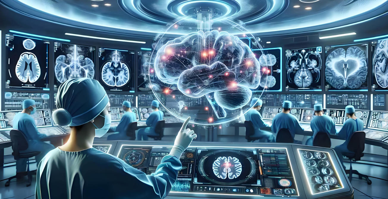
With the rapid advancement of modern healthcare, hospitals and surgical centers increasingly focus on precision diagnosis and minimally invasive treatment. Traditional medical imaging methods such as X-rays, CT scans, and 2D ultrasound are widely used but have clear limitations in providing real-time 3D spatial information, accurate surgical guidance, and high-precision navigation. TOF (Time-of-Flight) technology has emerged as a critical innovation, offering high-precision depth perception for medical imaging, lesion localization, surgical navigation, and intelligent diagnostics.
What is TOF Technology
TOF (Time-of-Flight) technology measures the distance and depth by calculating the time it takes for emitted light or infrared pulses to travel to a target and back to the sensor. Unlike conventional 2D cameras, TOF sensors can generate complete 3D depth maps of organs, tissues, or lesions, supporting volume measurement, lesion detection, motion tracking, and posture analysis.
Key advantages of TOF in healthcare include:
-
Non-contact measurement: No wearable devices or direct contact required, suitable for non-invasive or minimally invasive procedures.
-
High real-time performance: Captures depth data within milliseconds, ideal for real-time surgical guidance, monitoring, or rehabilitation feedback.
-
Versatile applications: Applicable in surgery, postoperative monitoring, smart health tracking, and remote medical care.

Limitations of Traditional Medical Imaging
Although X-rays, CT scans, and ultrasounds remain mainstream, they face challenges in complex or minimally invasive procedures:
-
Limited spatial information: 2D images cannot accurately show the 3D relationships between organs, lesions, and surrounding structures, making surgical planning less precise.
-
Suboptimal surgical planning: Narrow operative channels in minimally invasive surgery and limited visualization make it difficult to achieve optimal trajectories and angles.
-
Lower efficiency: Multiple 2D slices or views are required, consuming time and cognitive effort, especially for less experienced surgeons.
-
Difficulty capturing dynamic tissues: Organ motion due to breathing, heartbeat, or patient movement is not accurately reflected in traditional imaging, increasing surgical risks.
Key Value of TOF in 3D Medical Imaging and Minimally Invasive Navigation
With TOF, healthcare providers can overcome traditional imaging limitations, achieving more precise, safer, and intelligent diagnosis and treatment:
-
Real-time depth scanning: TOF systems emit light or infrared pulses and measure the return time to generate depth data within milliseconds, providing real-time organ and lesion monitoring without relying on CT or MRI.
-
Accurate volume and shape measurement: 3D point clouds enable precise calculation of organ or tumor volumes and shapes, guiding surgical resection while preserving healthy tissue.
-
Precise lesion, vessel, and structure localization: TOF provides 3D coordinates for planning minimally invasive paths, avoiding critical blood vessels, nerves, and functional areas.
-
Dynamic tissue tracking and real-time navigation: High refresh rates allow TOF to track organ motion from respiration or heartbeat, guiding surgical tools with high accuracy.
-
Assisted diagnosis and personalized treatment: 3D visual reports combined with AI automatically identify lesions, assess risks, and develop individualized surgical and rehabilitation plans.

Clinical Applications of TOF
-
Minimally invasive surgery navigation: Neurosurgery, cardiac surgery, laparoscopy, and arthroscopy use TOF for 3D path planning and real-time navigation, reducing incisions and tissue damage, and shortening recovery time.
-
Tumor localization and therapy: TOF helps locate tumors in real time, optimize surgery or radiotherapy targeting, and monitor tumor dynamics.
-
Vascular interventions and complex procedures: TOF assists in catheter navigation, stent placement, and vascular reconstruction, reducing complications.
-
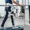
Postoperative rehabilitation and remote healthcare: Monitors joint mobility, posture recovery, and range of motion for personalized rehabilitation and remote care.
Challenges and Optimization
Despite its advantages, TOF faces challenges in clinical settings:
-
Soft tissue imaging difficulty: Low reflectivity of muscles, fat, or internal organs may cause signal attenuation or noise.
-
Light scattering and interference: Fluids, blood, moist tissues, or rapid motion can distort depth measurements.
-
Organ motion and environmental changes: Breathing, heartbeat, patient movement, and lighting interference may affect depth accuracy.
Optimization strategies include:
-
Using near-infrared wavelengths for better tissue penetration and signal stability.
-
Integrating AI and deep learning for noise reduction, multi-frame accumulation, dynamic compensation, and image reconstruction.
-
Multi-modal fusion with CT, MRI, ultrasound, or endoscopy to combine structural detail with real-time 3D depth data.
-
Enhancing TOF hardware to improve sensitivity, reduce noise, expand FOV, and adapt to complex surgical environments.

Future Trends
With AI, cloud computing, remote medical systems, and intelligent imaging platforms, TOF medical imaging will expand its applications:
-
TOF + AI intelligent diagnosis: Deep learning analyzes 3D point clouds for automatic lesion detection, segmentation, labeling, and risk assessment.
-
Personalized surgical planning: Patient-specific 3D models guide optimal minimally invasive paths and tool trajectories.
-
Intraoperative monitoring and navigation: Real-time updates of organ and lesion positions compensate for motion and guide robotic or manual instruments.
-
Remote healthcare and smart medical ecosystems: TOF data integrates with cloud platforms for teleconsultation, remote guidance, and rehabilitation monitoring.
-
Chronic disease management and health tracking: Long-term monitoring of posture, movement, respiration, and rehabilitation progress supports disease management and preventive care.
Conclusion
TOF technology provides high-speed, high-precision, non-contact 3D depth sensing that revolutionizes medical imaging, minimally invasive surgery, personalized treatment, postoperative care, and remote healthcare. Compared to traditional 2D imaging, TOF offers richer, more intuitive, and more reliable spatial information, greatly enhancing diagnostic, treatment, and surgical safety and efficiency. With AI, deep learning, multi-modal fusion, and intelligent healthcare systems, TOF medical imaging is becoming a foundational technology for precision medicine, smart surgery, and the future of healthcare ecosystems.
Okulo ™ C1 Precision RGB-Depth Imaging Camera: Cutting-Edge Visuals, State-Of-The-Art IToF Technology, And Seamless Hardware Integration

After-sales Service: Our professional technical support team specializes in TOF camera technology and is always ready to assist you. If you encounter any issues during the usage of your product after purchase or have any questions about TOF technology, feel free to contact us at any time. We are committed to providing high-quality after-sales service to ensure a smooth and worry-free user experience, allowing you to feel confident and satisfied both with your purchase and during product use.


Please upload banner from store admin blog pages